Pediatric cardiac surgery is one of the most demanding medical specialties. It needs to deal with delicate structures like tiny little hearts of neonates. The heart of the newborn child can weight even 20 grams, and fit on a human’s palm. Procedure on such a small heart brings a lot of difficulties.
Challenge
When the heart has only one abnormality, or it’s a well-known one, most of the doctors can base on their experience or the popular methods like CT scan and MRI to create an image of the abnormality The real challenge starts when the heart is tiny, the heart disease is rare, or it differs from other, typical cases. In all of those examples, even sub-millimeters can make a life-or-death difference. To increase the chance of survival, surgeons need to know as much as possible to decide whether to start the procedure or not and if so how to do it the best way.
In this case, Kordian, a 3-week old infant from Poland was suffering from the heart disease called traumatic aortic rupture. In this disorder aorta, the largest artery of the human body is torn or ruptured. This condition can be fatal, and for Kordian, there was also a threat to his life. Doctors along with Kordian’s mother, a person who didn’t have any knowledge about the disease, had to decide quickly about undertaking the procedure.
Solution
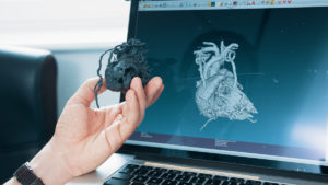
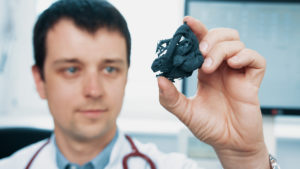
3D printing turned out to be a perfect solution for this problems. Cardiologists could print every heart, see the abnormality closely. They could even do a mock-up surgery before the real procedure. Unfortunately, while the most common FDM printers are good enough for everyday use, this time that technology couldn’t help. Doctors needed a solution that would provide surgical precision. They had to imitate every little vein and artery that surrounds the heart. For FDMs, veins were either too thin to print or, because of the needed supports, could be easily torn apart during post-processing.
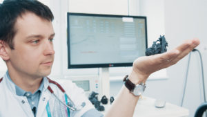
As it turned out, SLS technology meets all the requirements. Such SLS 3D-printed models can be used both for planning the cardiac surgery but also for interventional procedures, especially in complex and rare congenital heart diseases when the anatomy is always different and vary from patient to patient.
It helps to plan surgery and therefore make it safer, easier and shorter. Operations, where doctors practiced on 3D printed hearts, have a higher rate of success, which is leading to a better life of the patient.
Results implemented
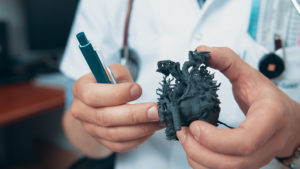
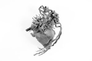
Surgeons decide to print Kordian’s heart to better plan an operation. With the rapture clearly visible on the SLS 3D printed model, the minimal size of the heart wasn’t a problem. It helped to prepare for the complicated operation. They connected raptured fragments of the aorta. Seeing the model made it easier for Kordian’s mother to understand how serious his condition was, and how the procedure will proceed. Thanks to that, the consent to the operation was more conscious than if she only saw the defect on the screen.
Today Kordian is 18 months old. His happy face and positive attitude toward new people don’t reveal that over a year ago this boy could not live.
SLS 3D printing helped with:
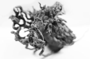
Creating a 1:1 heart model of a neonate, that maintain structure and relation of different anatomical structures like veins, trachea or arteries, not possible to achieve in other technologies,
Helping students to learn about various heart diseases in the most visual way Planning and mocking upcoming procedures, what leads to a higher survival rate of patients,
Explaining the heart diseases to parents, who are not aware of heart abnormalities in general.